Home
/ Bone Cross Section View - Classification Of Tissues Histology Ppt Download / Smartdraw includes 1000s of professional healthcare and anatomy chart templates that you can modify and make your own.
Bone Cross Section View - Classification Of Tissues Histology Ppt Download / Smartdraw includes 1000s of professional healthcare and anatomy chart templates that you can modify and make your own.
Bone Cross Section View - Classification Of Tissues Histology Ppt Download / Smartdraw includes 1000s of professional healthcare and anatomy chart templates that you can modify and make your own.. The outside of a bone is covered in a thin layer of dense irregular connective tissue called the periosteum. They are obtained by taking imaginary slices perpendicular to the main axis of organs, vessels, nerves, bones, soft tissue, or even the entire human body. Label the membrane that lines the cavity and the membrane that covers the outside surface. Cortical area (ca, mm 2) was calculated as the area of compact bone (defined by threshold values) within a given cross section. In a cross section of a bone we can see two types of bone tissue:
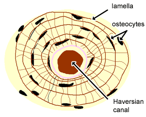
A, axial section through the roots of the maxillary teeth. When the bone section is viewed under transmission electron microscope, it is possible to see collagen that makes up most of the organic matrix. The periosteum contains many strong collagen fibers that are used to firmly anchor tendons and muscles to the bone for movement. (b) in this micrograph of the osteon, you can see the concentric lamellae around the central canals. Looking at a bone in cross section, there are several distinct layered regions that make up a bone.
Bone Section With Skin Doctor Stock from m.psecn.photoshelter.com Bone structure cross sectional view of the femoral head, bone, bone structure cross sectional view of the femoral head. This file is licensed under the creative commons attribution 3.0 unported license.: Do not color the articular cartilage; Select different colors for the structures and bone areas in column b, and use them to color the coding circles and corresponding structures on the figure diagrams. Drag and drop upload, viewable on any device. We can see there are two layers of compact bone here. (b) in this micrograph of the osteon, you can see the concentric lamellae around the central canals. The result is called a front section, or front view in section, since it replaces the front view in the drawing.
The spongy and compact bone tissue in the cross section of a skull bone.
Whereas a long bone has only one layer of compact bone (see fig 1). The periosteum contains many strong collagen fibers that are used to firmly anchor tendons and muscles to the bone for movement. Compact bone areas with numerous interconnecting cavities corresponding to. This photograph shows a section through a marrow space within a bone. They are obtained by taking imaginary slices perpendicular to the main axis of organs, vessels, nerves, bones, soft tissue, or even the entire human body. This file is licensed under the creative commons attribution 3.0 unported license.: 3d illustration of a cross section of the human eye with explanations and inscription In addition, students can see the osteoid tissue, which is uncalcified matrix. This is a short tutorial using blender 2.8 that shows how to create a bone cross section and using images to create the textures.hope you enjoy and please su. Do not color the articular cartilage; (b) in this micrograph of the osteon, you can clearly see the concentric lamellae and central canals. (b) in this micrograph of the osteon, you can clearly see the concentric lamellae and central canals. Label the membrane that lines the cavity and the membrane that covers the outside surface.
(b) in this micrograph of the osteon, you can clearly see the concentric lamellae and central canals. (b) in this micrograph of the osteon, you can see the concentric lamellae around the central canals. Bone structure cross sectional view of the femoral head, bone, bone structure cross sectional view of the femoral head. They are obtained by taking imaginary slices perpendicular to the main axis of organs, vessels, nerves, bones, soft tissue, or even the entire human body. Learn vocabulary, terms, and more with flashcards, games, and other study tools.
Cartilage Bone Ossification The Histology Guide from www.histology.leeds.ac.uk The osteon has blood vessels and bone cells, things vital for the survival of the bone. We can see there are two layers of compact bone here. In this short video i use blender 2.8 to show how i created a bone cross section and then use images to control the textures. Do not color the articular cartilage; For example, if there was a hollow cavity, or internal features, or anything that you can't see from the outside of the part, this view can come in handy. Smartdraw includes 1000s of professional healthcare and anatomy chart templates that you can modify and make your own. Looking at a bone in cross section, there are several distinct layered regions that make up a bone. Foot bone anatomy x ray 12 photos of the foot bone anatomy x ray foot bone anatomy x ray, bone, foot bone anatomy x ray.
You may do so in any reasonable manner, but not in any way.
Foot bone anatomy x ray 12 photos of the foot bone anatomy x ray foot bone anatomy x ray, bone, foot bone anatomy x ray. This file is licensed under the creative commons attribution 3.0 unported license.: This photograph shows a section through a marrow space within a bone. Label the membrane that lines the cavity and the membrane that covers the outside surface. They are obtained by taking imaginary slices perpendicular to the main axis of organs, vessels, nerves, bones, soft tissue, or even the entire human body. As figures shown, the cutting plane is a frontal plane and appears as a line in the top view. Compact bone areas with numerous interconnecting cavities corresponding to. Cross section of a human bone showing bone marrow, spongy bone and blood vessels. Smartdraw includes 1000s of professional healthcare and anatomy chart templates that you can modify and make your own. Drag and drop upload, viewable on any device. The periosteum contains many strong collagen fibers that are used to firmly anchor tendons and muscles to the bone for movement. A, axial section through the roots of the maxillary teeth. Color the bone tissue gold.
Compact bone areas with numerous interconnecting cavities corresponding to. Color the bone tissue gold. The periosteum contains many strong collagen fibers that are used to firmly anchor tendons and muscles to the bone for movement. They are obtained by taking imaginary slices perpendicular to the main axis of organs, vessels, nerves, bones, soft tissue, or even the entire human body. Select different colors for the structures and bone areas in column b, and use them to color the coding circles and corresponding structures on the figure diagrams.
A Layout Of Bone Marrow In A Cross Sectional View Of A Tubular Bone Download Scientific Diagram from www.researchgate.net The result is called a front section, or front view in section, since it replaces the front view in the drawing. 3d illustration of a cross section of the human eye with explanations and inscription As the names suggest compact bone looks compact and the spongy bone looks like sponges. Cross section of a human bone showing bone marrow, spongy bone and blood vessels. You may do so in any reasonable manner, but not in any way. Sectional views often replace standard views. Cross section area is an area of an object if you view it as a 2d object. Learn vocabulary, terms, and more with flashcards, games, and other study tools.
(b) in this micrograph of the osteon, you can clearly see the concentric lamellae and central canals.
Cross section through an model of an normal upper right femur or leg bone. They are obtained by taking imaginary slices perpendicular to the main axis of organs, vessels, nerves, bones, soft tissue, or even the entire human body. We can see there are two layers of compact bone here. Cross section of a femur bone showing the anatomical structure including cancellous bone and marrow. The spongy and compact bone tissue in the cross section of a skull bone. Bone on side of the foot This is a short tutorial using blender 2.8 that shows how to create a bone cross section and using images to create the textures.hope you enjoy and please su. As the names suggest compact bone looks compact and the spongy bone looks like sponges. In this short video i use blender 2.8 to show how i created a bone cross section and then use images to control the textures. Bone structure cross sectional view of the femoral head, bone, bone structure cross sectional view of the femoral head. This file is licensed under the creative commons attribution 3.0 unported license.: Cancellous bone has large open spaces (marrow spaces) and plates of bone called trabeculae. The outside of a bone is covered in a thin layer of dense irregular connective tissue called the periosteum.
(b) in this micrograph of the osteon, you can clearly see the concentric lamellae and central canals bone cross section. The result is called a front section, or front view in section, since it replaces the front view in the drawing.

